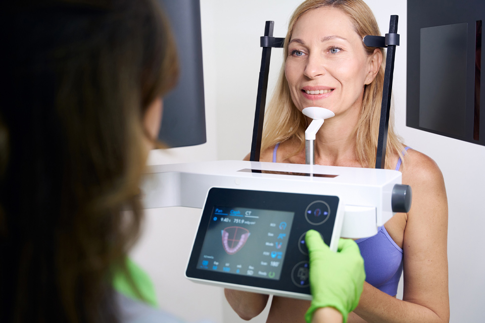
For precise diagnostics and safe treatment planning, we use three-dimensional imaging with CBCT (Cone Beam Computed Tomography) at our practice in Freienstein. Unlike conventional two-dimensional X-rays, CBCT shows the entire jaw, teeth and surrounding structures in three dimensions — with exceptional detail and comparatively low radiation exposure.
3D diagnostics gives our clinical team a complete, accurate picture of your individual anatomy before any treatment begins. This means less guesswork, fewer surprises during procedures, and consistently better outcomes for our patients. Our approach is rooted in a broader metal-free dentistry approach that prioritises biocompatibility and minimal intervention at every stage.
Three-dimensional CBCT imaging is particularly valuable in a wide range of clinical situations. We routinely use it for:
In each case, our goal is to act only on a thoroughly verified diagnosis — so that treatment is targeted, minimally invasive and as effective as possible.
Is the radiation exposure from a CBCT scan dangerous?
No. A CBCT scan has significantly lower radiation than a conventional medical CT scan — roughly equivalent to 2–5 standard dental X-rays, depending on the region scanned and the device settings used. We always apply the ALARA principle (As Low As Reasonably Achievable) and only recommend a CBCT scan when the clinical benefit clearly outweighs the minimal radiation dose.
How long does a CBCT scan take?
The actual scan takes between 10 and 40 seconds. Including positioning and preparation, the whole process typically takes no more than 10 minutes. The resulting data is immediately available for evaluation on our diagnostic software.
Will I need a CBCT scan before every treatment?
No. We only use 3D diagnostics when it provides clinically relevant information that cannot be obtained from a conventional X-ray. For routine check-ups and many standard treatments, a 2D X-ray is perfectly sufficient.
Can CBCT diagnostics be combined with other biological treatments?
Yes. CBCT findings frequently guide other biological therapies at our practice. For example, identifying post-extraction healing deficits may lead us to recommend PRF therapy to support healing, or targeted treatment of FDOK/NICO lesions confirmed by the scan.